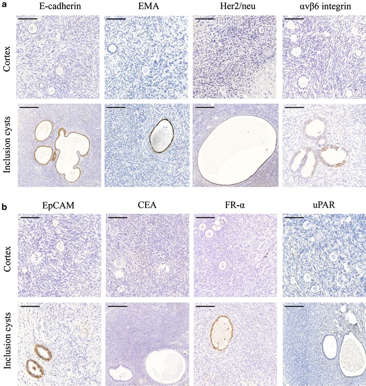
Fig. 1.
a Immunohistochemical expression of E-cadherin, EMA, Her2/neu and αvβ6 integrin in ovarian cortices and inclusion cysts. Stromal cells stained negative, but E-cadherin, EMA, Her2/neu and αvβ6 integrin showed expression at the epithelial cells of inclusion cysts. Scale bars in the upper panel represent 100 μm and scale bars in the lower panel represent 200 μm. b Immunohistochemical expression of EpCAM, CEA, FR-α and uPAR in ovarian cortices and inclusion cysts. Stromal cells stained negative, but EpCAM and FR-α showed expression at the epithelial cells of inclusion cysts. Scale bars in the upper panel represent 100 μm and scale bars in the lower panel represent 200 μm

