Fig. 2.

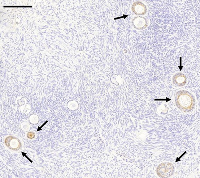
Immunohistochemical staining of E-cadherin showed moderate expression in the granulosa cells of primary follicles in the ovarian cortex (arrows). Scale bar represents 200 μm

Immunohistochemical staining of E-cadherin showed moderate expression in the granulosa cells of primary follicles in the ovarian cortex (arrows). Scale bar represents 200 μm