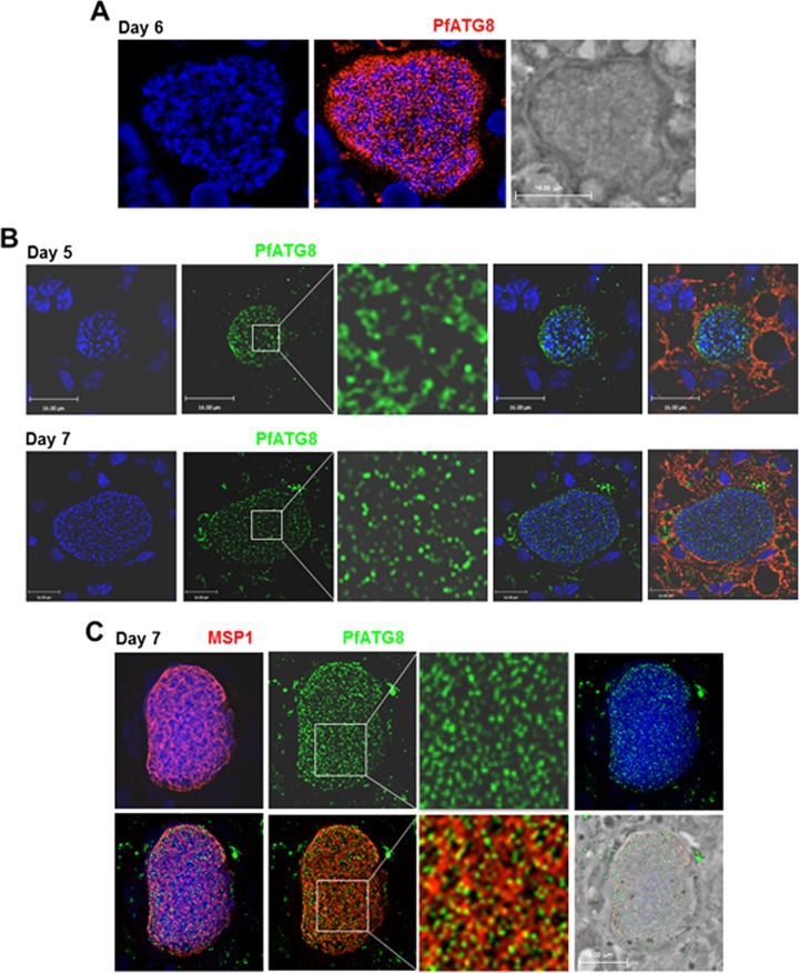
FIG 1 .
Expression of ATG8 by liver forms of Plasmodium falciparum in a humanized mouse. Immunofluorescence assays on mouse liver sections containing P. falciparum-infected human hepatocytes using anti-PfATG8 antibodies (red [A] or green [B]) at the indicated days p.i. In panel B, cells were counterstained with Evans blue. In panel C, the parasites were immunostained with antibodies against PfATG8 (green) and MSP1 for the plasma membrane of hepatic merozoites (red). DAPI (blue) was used for staining nuclei in all panels. The vast majority of the PfATG8 signal was associated with the PV. Some surrounding liver cells were also reacting with the anti-PfATG8 antibody: nonspecific background staining is inherent in liver tissue due to cell necrosis and high intracellular protein content. Bars, 16 μm.

