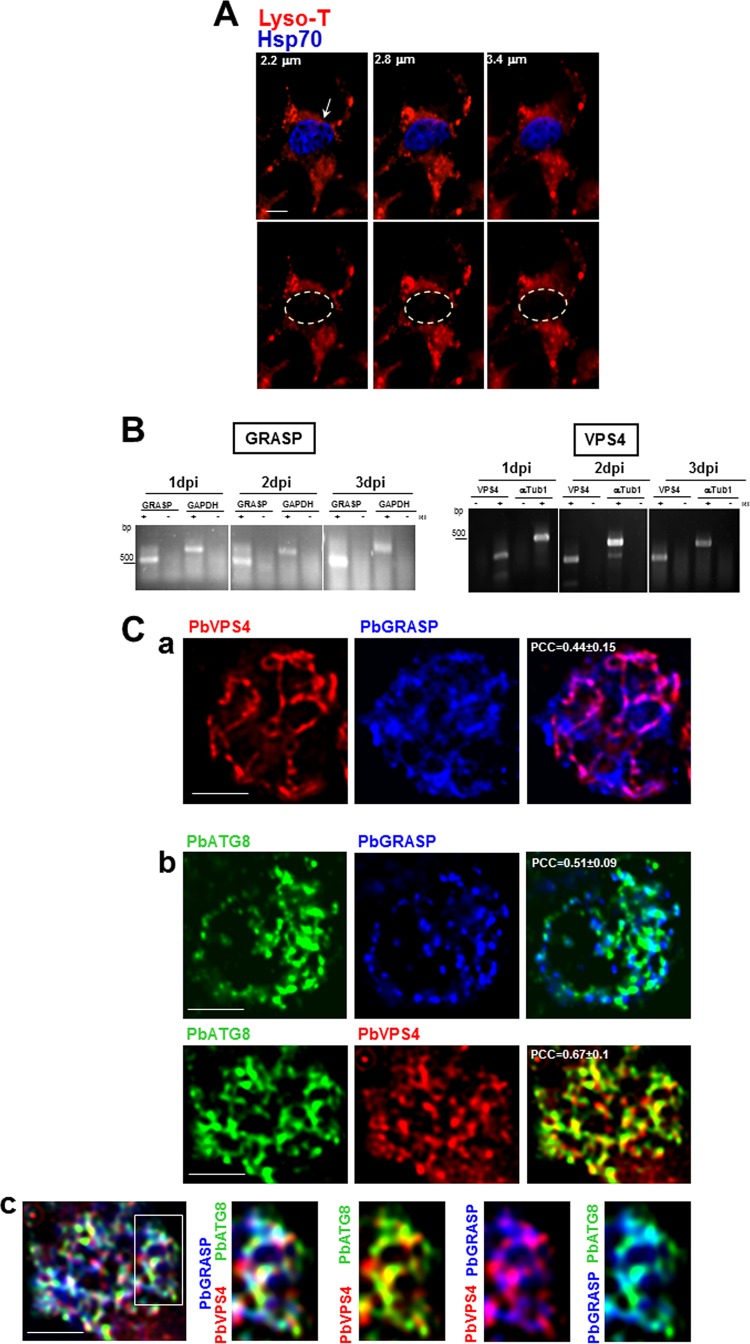
FIG 3 .
Coassociation of PbATG8 with GRASP- and VPS4-positive structures. (A) Fluorescence assays on P. berghei-infected cells stained with LysoTracker (red) 40 h p.i. Parasites were identifiable by immunolabeling for Hsp70 (blue). Three optical z-slices of a PV are shown. Bar, 5 µm. (B) Transcriptional profiles of GRASP and VPS4 in P. berghei liver stages. Expression in liver-stage parasites at 1, 2, and 3 days p.i. was assayed by RT-PCR. To verify the absence of genomic DNA contamination, RT-PCRs were set up in duplicate with (+) and without (−) reverse transcriptase (RT). GAPDH or α-tubulin (αTub1) was used as an internal control. PCCs were calculated from 3 independent assays. (C) Double (a and b) or triple (c) IFA on P. berghei-infected cells 30 h p.i. using antibodies against PbATG8, PbGRASP, and PbVPS4. PCCs were calculated from 3 independent parasite preparations (n = 17 to 26 PVs). Bars, 10 µm.

