Figure 1.

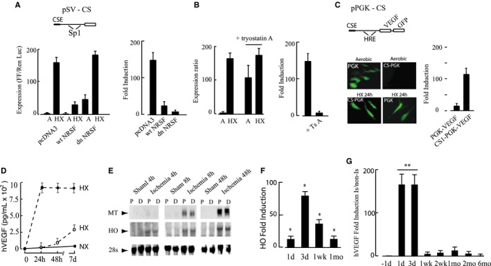
Mechanism of conditional silencing. A, C2C12 skeletal myocytes were transfected with pSV40‐CS‐Luc (pSV‐CS) (2 μg) and pcDNA, pNRSF, or pdnNRSF (2 μg), with Renilla luciferase as internal control and exposed to normoxia (21% O2) or hypoxia (0.5% O2). In the promoter diagram, CSE indicates conditional silencer element, and Sp1 depicts the positions of two Sp1 transcription factor binding sites in the SV40 promoter. B, C2C12 skeletal myocytes were transfected with pSV‐CS and subjected to aerobic or hypoxic incubation as in A; parallel plates were treated for 12 hours with trichostatin A (250 ng/mL) or vehicle. C, Adeno‐associated virus (AAV) shuttle plasmids expressing green fluorescent protein (GFP) and vascular endothelial growth factor (VEGF) phosphoglycerate kinase (PGK) directed by the PGK promoter or conditionally silenced PGK promoter (CS) were transfected into C2C12 skeletal myocyte and cultured for 48 hours under normoxia followed by 24 hours’ hypoxia. GFP was visualized by the use of fluorescence microscopy, and human VEGF (hVEGF) by the use of ELISA as described in Methods. D, C2C12 myocytes were infected with dsAAV1‐CS‐hVEGF (closed circles) or ssAAV9‐CS‐hVEGF (open circles) as described in Methods. Cultures were exposed to hypoxia (HX) or normoxia (NX) as indicated and samples of culture medium taken at the indicated times. Secreted hVEGF was measured by ELISA and values indicate rate of hVEGF production (pg/mL per 24 hours) for each condition. E, Mouse hindlimbs were made ischemic or subjected to sham operation as described in Methods. Mice were killed at 4, 8, 24, and 48 hours; the thigh muscles removed and cut into 2 longitudinal pieces proximal (P) and distal (D) to the femoral artery (FA) ligature. RNA was extracted and analyzed by Northern blot. F, Transcript levels of the HO gene in proximal adductor muscles containing the excised FA were quantified by RT‐PCR. G, Hindlimbs of BALB/c mice were injected with 1×10¹¹ VP AAV‐CS‐VEGF, 5 days before implementing ischemia. Adductor muscle containing the injection sites was harvested and the transcript levels of hVEGF quantified by RT‐PCR. All results are mean±SEM, n=4; for F and G, *P<0.05; **P<0.01 by Student t test.
