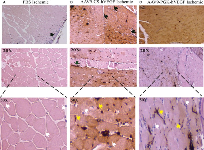
Figure 4.

Human vascular endothelial growth factor (hVEGF) immunostain. A through C, Mouse hindlimbs were preinjected with phosphate buffered saline (PBS) or the indicated vectors (1011 VPs) 5 days before femoral artery excision as described in Methods. Lower thigh and calf muscles were harvested on day 3, dissected, fixed, and stained with anti‐hVEGF antibody, and microscopic images were obtained in a semiblinded manner as described in Methods. Black arrows indicate border of positive and negative hVEGF staining; white and yellow arrows depict capillaries and inflammatory cell infiltrates, respectively, that may be negative (blue) or positive (dark brown) for hVEGF. AAV indiactes adeno‐associated virus; CS, conditionally silenced; PGK, phosphoglycerate kinase.
