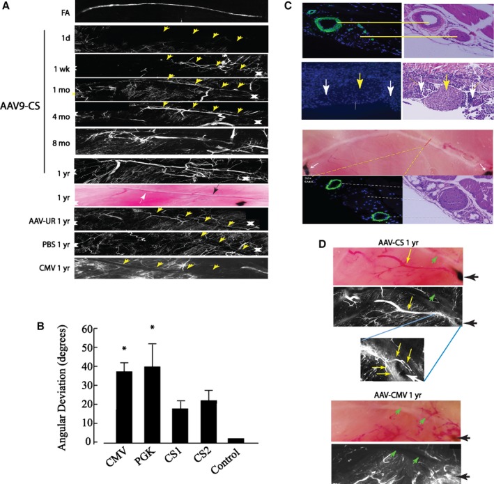
Figure 6.

Femoral artery (FA) regeneration by conditionally silenced (CS) adeno‐associated virus (AAV)‐human vascular endothelial growth factor (hVEGF). A, Composite DiI images show vessel regeneration in AAV9‐CS‐hVEGF–treated limbs. Vessels regenerate along the femoral tract (yellow arrowheads) over the course of 1 year after FA excision. Progressive vessel regeneration is not seen in limbs treated with AAV‐CMV‐hVEGF, AAV‐PGK‐hVEGF, or phosphate buffered saline (PBS). B, Composite DiI images were analyzed by using Velocity software to measure the angular deviation of regenerated vessels from the path of the femoral nerve as described in Methods (*P<0.05; n=4 limbs×10 vessels per limb=40 vessels per group). C, Top panel, anti‐smooth muscle actin (SMA)– and DAPI‐ (left) and hematoxylin and eosin (H&E)– (right) stained sections containing the normal FA and femoral nerve; second panel, the same location immediately after hindlimb ischemia surgery; yellow arrow indicates femoral nerve; white arrows indicate region of FA excision. Bottom panels, top: light field image of hindlimb muscle with AAV9‐CS‐hVEGF treatment at 10 months after FA excision. Anti‐SMA and H&E stains confirm arterial regeneration within the femoral tract (bottom panels). D, Light field (top) and DiI image (middle) of AAV‐CS‐hVEGF treated hindlimb at 1 year after ischemia surgery; inset (bottom) shows higher magnification of vessels skirting the sutures. E, Same as (D) except treatment with AAV9‐CMV‐hVEGF. CMV indicates cytomegalovirus; PGK, phosphoglycerate kinase.
