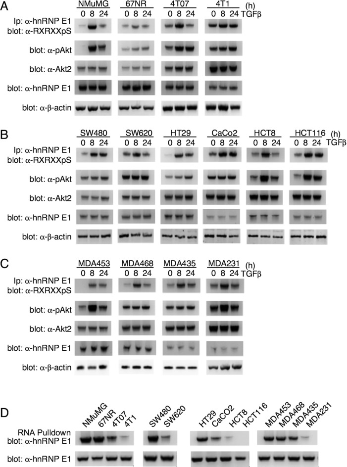
Figure 7.
pAkt2 and p-hnRNP E1 are significantly upregulated in metastatic cancer lines. (A) Immunoblot analysis of p-hnRNP E1, p-Akt, Akt2 and hnRNP E1 in NMuMG cells and the 4T01 metastatic progression series. 67NR cells are tumorigenic and not metastatic, 4TO7 are metastatic, yet the metastases do not form secondary tumors and 4T1 cells are metastatic and form secondary tumors. Akt2 activation is measured by assaying phosphorylated Ser474 (p-Akt2), and p-hnRNP E1 levels are analyzed through a combinatorial α-hnRNP E1 immunoprecipitation followed by α-Akt2 phospho-substrate immunoblotting. The immunoblot data presented are typical of three independent experiments demonstrating similar levels of protein expression levels. (B and C) Human colon (B) and breast (C) cancer cell lines of varying metastatic potential were analyzed as in (A). We utilized the progression series analysis as a means of correlating metastatic potential to human tumor cell lines. The immunoblot data presented are typical of three independent experiments demonstrating similar levels of protein expression levels. (D) To demonstrate the effect that p-hnRNP E1 : hnRNP E1 ratios play on interaction with a consensus motif element, we performed an RNA pulldown assay followed by α-hnRNP E1 immunoblot analysis. When compared with total hnRNP E1 levels, a significant loss of interaction is noted in highly aggressive cell lines.

