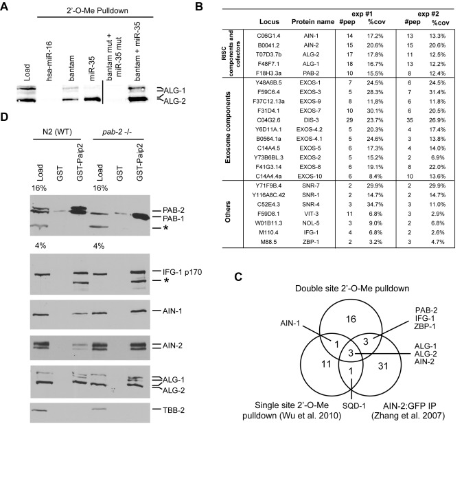
Figure 1.
PAB-1 and PAB-2 interact with miRISC in Caenorhabditis elegans embryos. (A) ALG-1/2 western blot analysis of 2′-O-Me pull-downs. 5′ Biotinylated 2′-O-Me oligos containing miR-35, CeBantam or both sites were used to pull down the miRISC WT strain (N2) embryonic extract. ALG-1 and ALG-2 were detected using near-infrared fluorescent western blot (LiCOR). (B) Proteins identified in MuDPIT analyses of a dual site (CeBantam + miR-35) 2′-O-Me pull-down. Only proteins which were detected in two independent purifications and were absent from pull-downs with mutated binding sites are included here. For each protein, the number of peptides (#pep) found in each experiment and the coverage (%cov) of these peptides for the full-length protein are indicated. (C) Venn diagram comparing detected interactions with the dual site (CeBantam + miR-35) pull-down to single-site RISC pull-down, and AIN-2-GFP IP. (D) A GST pull-down using either GST or GST-PAIP2 was performed either on WT (N2) or mutant pab-2(0) (ok1851) embryonic extract in the presence of 0.1 ng/μl RNAse A. The bound proteins were analyzed on SDS-PAGE and detected by western blot.*: non-specific band.

