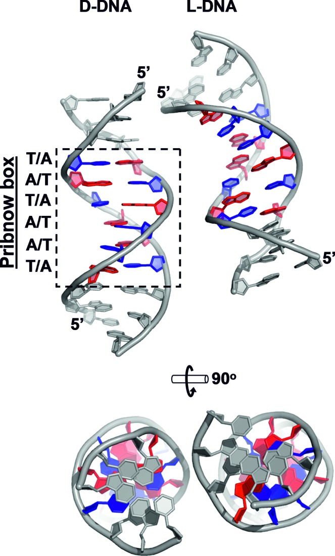
Figure 2.

Crystal structure of a DNA duplex containing the Pribnow box consensus promoter sequence (crystal structure 4, space group Pbca, resolution 1.65 Å). Left: asymmetric unit, containing one complete D-DNA duplex. Right: L-DNA symmetry mate. Pribnow box residues are coloured blue (thymine) and red (adenine). All other residues (i.e. all non-Pribnow box residues) are coloured grey.
