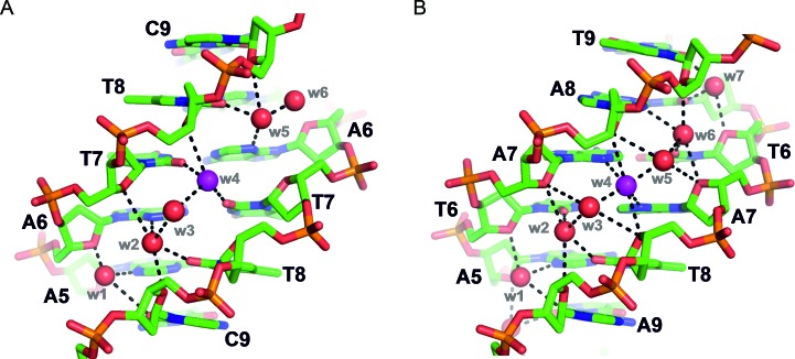
Figure 4.
Comparison of minor groove water structure and hydrogen bonding between: (A) a non-racemic model DNA duplex (Dickerson–Drew dodecamer, in space group P212121, PDB ID: 1BNA (35)) and (B) a racemic Pribnow box duplex (in space group P21/c [structure 2 (A) in Table 1]). The water structure of the minor groove is well-conserved overall between these two duplexes—one extra water molecule appears in the Pribnow box duplex—despite the difference in sequence. In the parts shown, minor groove hydrogen bond acceptors may be adenine N3 atoms, or thymine or cytosine O2 carbonyl groups. See for example w4 shown in magenta, hydrogen bonded to thymine-7 bases in (A) and to adenine-7 bases in (B).

