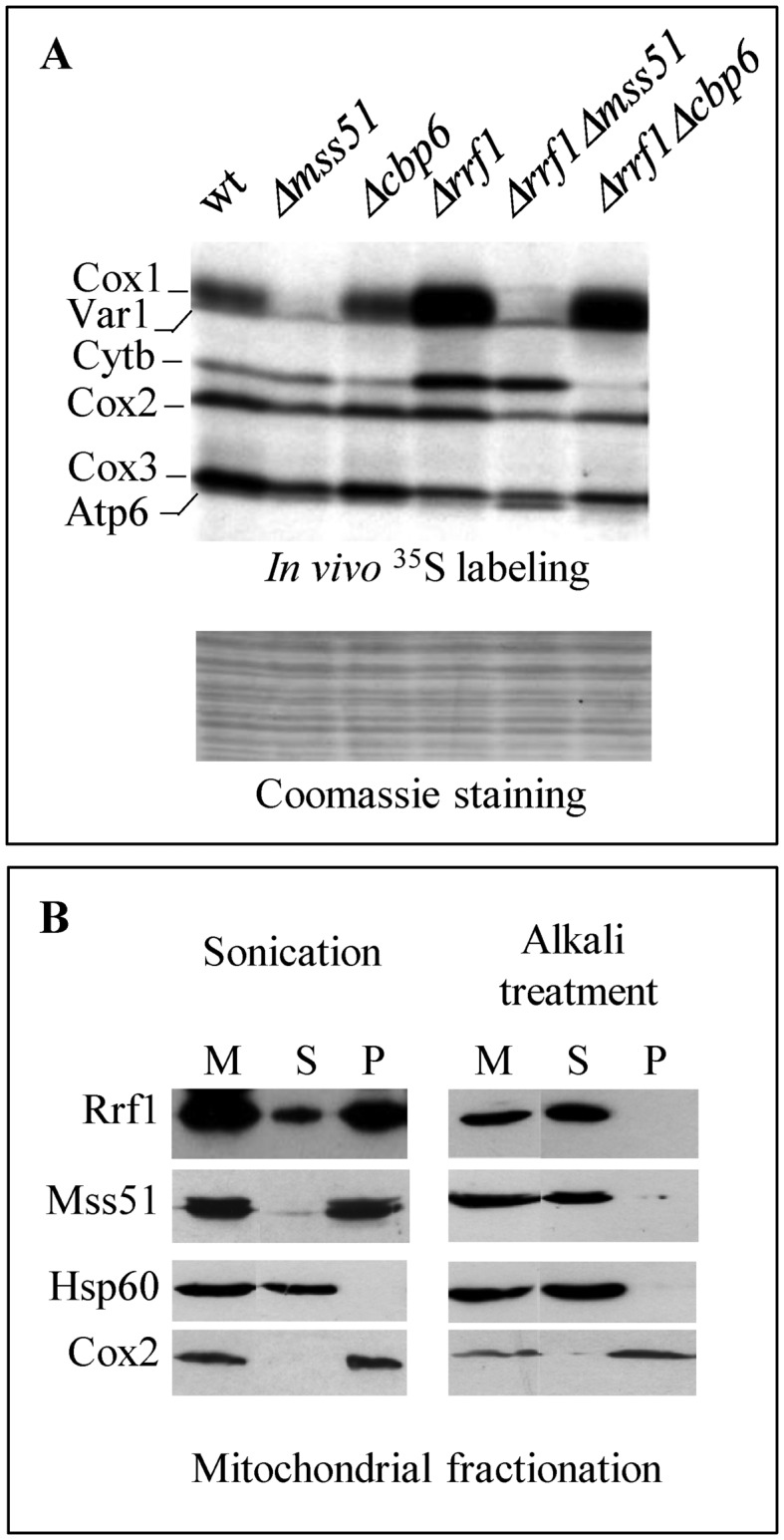
Figure 5.

Genetic interactions and localization of Rrf1 and the translational activators. (A) In vivo [35S] incorporation as in Figure 2A. (B) Mitochondria were purified from cells producing Mss51-c-Myc. Mitochondrial proteins (M) were either sonicated (left) or alkali treated (right) and after centrifugation the soluble proteins were recovered in the supernatant (S) whereas the membrane-associated or membrane-spanning proteins remained in the pellet (P). Proteins were analyzed by Western blotting with antibodies recognizing Rrf1 or the epitope c-Myc. The soluble matrix protein Hsp60 and the integral mitochondrial membrane protein Cox2 were used as controls.
