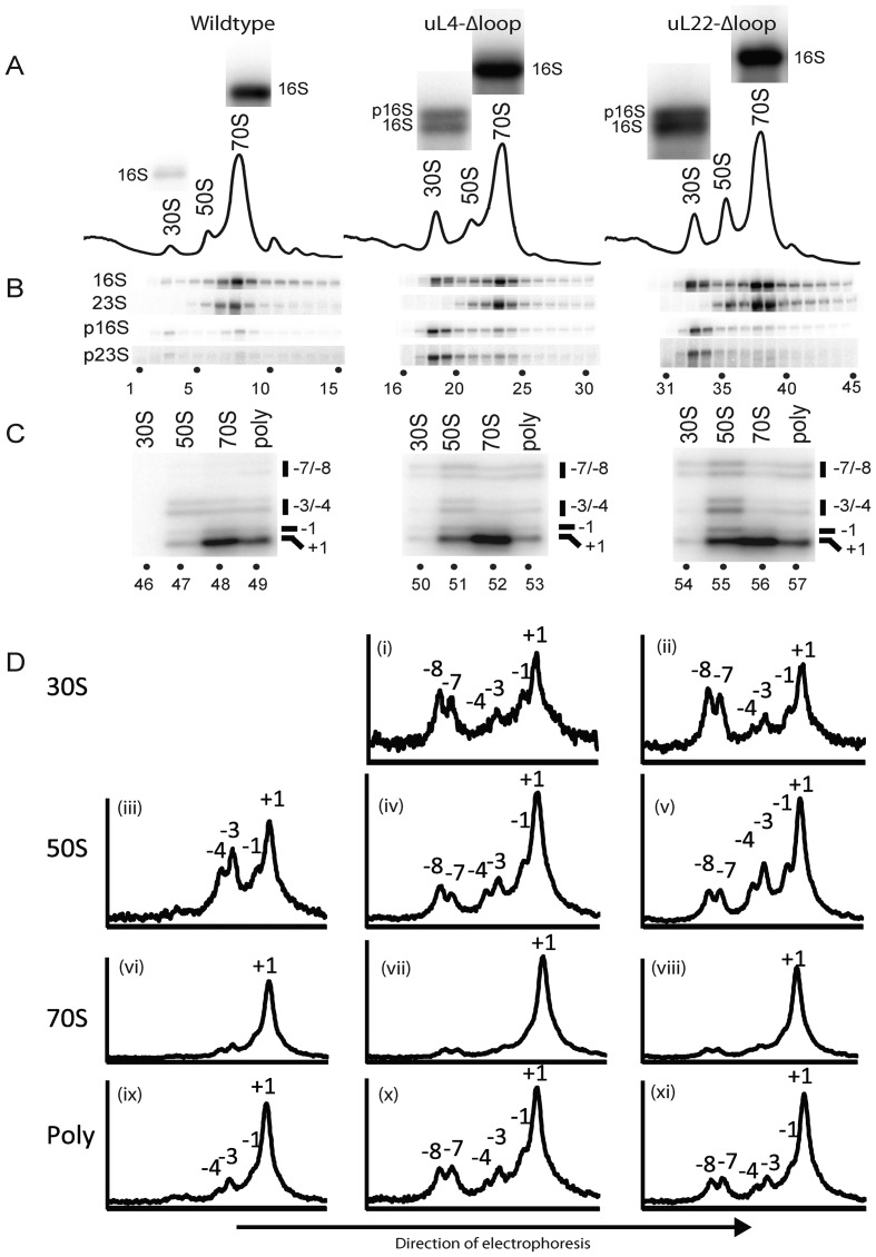
Figure 4.
Analysis of mutant and parent ribosomes. Cultures of the indicated strains were grown at 37°. (A) Sucrose gradient profiles. Ribosomes were fractionated on sucrose gradients in Buffer A1 (associating conditions). The bands shown above each peak are explained in the legend for part of the figure (B). (B) Northern analysis of gradient fractions containing ribosomal peaks. RNA was prepared from each sucrose gradient fraction and used for a northern blot, which was probed with oligonucleotides specific for precursor 16S (p16S), precursor 23S (p23S) and mature 16S and 23S rRNA molecules (see text, Supplementary Table S1 and Supplementary Figure S1 for more information about the probes). The bands from the hybridization with the 16S probe to RNA from the 30S and 70S peak fractions were enlarged and shown above each peak in part A. Note that RNA from the 30S peak from the two mutants, but not from the wild-type, contains two types of rRNA hybridizing to the 16S probe. The top bands are precursor 16S (also called 17S) and the bottom bands are mature 16S rRNA. (C) 5′ end mapping of 23S-type rRNA in sucrose gradient peaks. A second sucrose gradient was run on the ribosome preparations from each of the three strains and 84 fractions were collected from each gradient. Fractions from the 30S, 50S, 70S and polysome regions were then pooled and RNA from each pool was used for primer extension using a primer complementary to nucleotides 72–89 of mature 23S rRNA. The products were fractionated on gels and imaged on a phosphoimager. (D) Quantification of primer extension results shown in C. The pixel density along each lane of the gel image in C was digitized using ImageJ and plotted. The ordinates show the relative pixel density at different points of the gel. Note that the ordinates for different plots (corresponding to different lanes) are not the same. The plots serve only to show the distribution within a lane. The peaks of 23S rRNA with mature and 5′ extensions of 1, 3, 4, 7 and 8 nucleotides are indicated. Panel numbers are referred to in the text.

