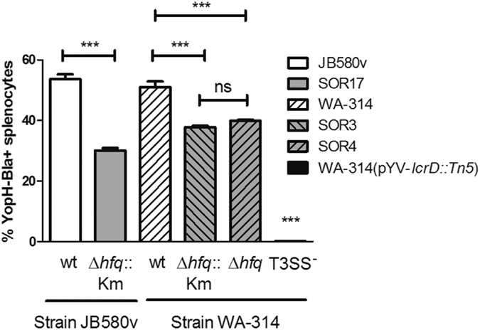
Figure 6. Hfq promotes translocation of YopH into splenocytes.

Bacterial strains carrying a YopH-β-lactamase fusion were grown for 90 min at 37 °C in BHI prior to infection of isolated mice splenocytes at a MOI of 20. After 75 min of infection, cells were stained with the green fluorescent substrate CCF4-AM for 90 min. Blue fluorescence (resulting from substrate cleavage by injected YopH-Bla) and green fluorescence of splenocytes were measured by flow cytometry. Results represent the mean percentage of blue cells and standard deviation of duplicate infections and are representative of two independent experiments. Significance was calculated with One-way ANOVA (P < 0.001) with post-hoc Bonferroni’s Multiple Comparison test (***P ≤ 0.001; ns, not significant P > 0.05). The CFUs in the inoculum were determined by plating and were as follows: JB580v, 1.7 × 10⁷; SOR17, 1.9 × 10⁷; WA-314, 3.9 × 10⁷; SOR3, 7.2 × 10⁷; SOR4, 5.9 × 10⁷.
