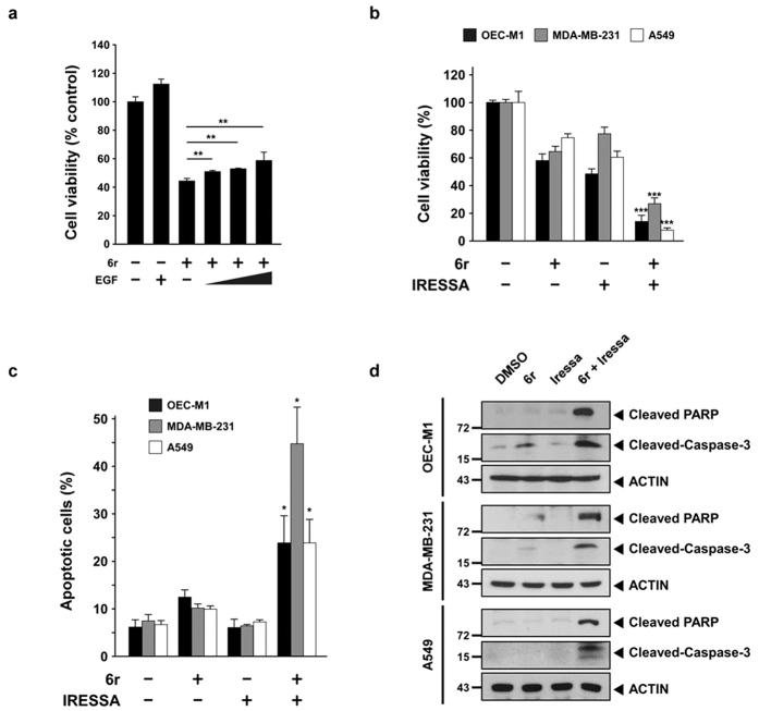
Figure 7. Cotreatment with 6r and Iressa enhances cancer cells apoptosis.
(a) OEC-M1 cells were pretreated with or without 20 μM 6r for 1 h and subsequently incubated with 100 ng/mL EGF for an additional 72 h. Cell viabilities were determined by a methylene blue dye assay. The bars represent the relative mean survival of four independent wells ± SD. Differences were considered statistically significant at *P < 0.05, **P < 0.01, and ***P < 0.001. (b) The cells were treated with 6r alone or in combination with Iressa for 72 h. Cell viabilities were determined by a methylene blue dye assay. In the OEC-M1 cells, 20 μM 6r and 10 μM Iressa were used. For the MDA-MB-231 cells, 15 μM 6r and 15 μM Iressa were used. Moreover, 40 μM 6r and 27 μM Iressa were used in the A549 cells. (c) After 24 h of treatment with 6r or Iressa or the 6r–Iressa combination, cell apoptotic rates were evaluated with annexin V/propidium iodide double staining. The data represent the mean ± SD of three independent experiments. Differences were considered statistically significant when *P < 0.05, **P < 0.01, and ***P < 0.001. (d) The cells were treated with 6r, Iressa, or the 6r–Iressa combination for 24 h; cleaved PARP and caspase-3 were assessed through Western blotting.

