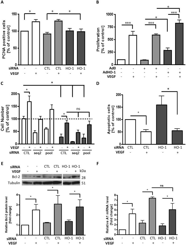
Figure 1. siRNA-mediated HO-1 depletion abrogates VEGF-induced HUVEC proliferation.
(A) HUVEC were transfected with control siRNA (CTL) or HO-1-specific siRNA prior to culture in the presence or absence of VEGF (25 ng/ml) for 48 h. Proliferation was quantified by flow-cytometric analysis of PCNA staining. (B) HUVEC were left uninfected or infected with the Ad0 control adenovirus or AdHO-1 (multiplicity of infection (MOI) 100 ifu/cell). EC were then cultured in the absence or presence of VEGF for 48 h and proliferation quantified by BrdU ELISA. (C) HUVEC were treated with vehicle alone (dotted line) or transfected with control siRNA (CTL) or HO-1 specific siRNA (seq2 or 4 pooled sequences (pool)). Cells were cultured in M199 medium/10% FCS (white bars) or serum-starved in M199/0.1% BSA in the absence or presence of VEGF (25 ng/ml) for 48 h (dark bars). Cell number was assessed by MTS-assay. (D) HUVEC were transfected with control (CTL) or pooled HO-1 siRNA oligos (HO-1) as above prior to culture with or without VEGF and serum starvation. Sub-G1 apoptotic EC were identified by propidium iodide staining and quantified by flow-cytometry, with data expressed as a percentage of the apoptosis seen in control siRNA-treated serum-starved cells. (E,F) HUVEC were transfected with control or pooled HO-1 siRNA (HO-1) and cultured in the absence or presence of VEGF for 48 h, with (E) Bcl-2 protein analysed by immunoblotting and quantified by densitometry, and (F) A1 mRNA quantified by qRT-PCR. Data are presented as mean ± SEM (n ≥ 4 experiments), *p < 0.05, **p < 0.01, ***p < 0.001, ns = not significant.

