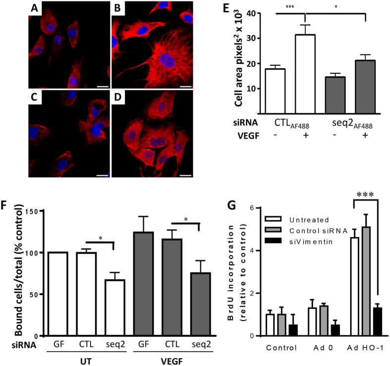
Figure 8. Vimentin filament assembly and EC adhesion is impaired by HO-1-depletion.
HUVEC, transfected with (A,B) CTLAF488 or (C,D) HO-1 siRNA seq2AF488, were seeded onto gelatin-coated coverslips in the absence (UT) or the presence of 25 ng/ml VEGF for 16 h, prior to fixation and vimentin staining. Nuclei were stained with Draq5. Immunofluorescence was analyzed by confocal microscopy. Representative images are shown, 63x magnification, bar = 50 μm. (E) Cell area was quantified using ImageJ (n = 15). (F). HUVEC treated with geneFECTOR (GF) alone or transfected with control (CTL) or HO-1 siRNA (seq2) were labelled with 5-chloromethylfluorescein diacetate (6.25 μM) and seeded onto gelatin-coated 96-well plates in the absence (UT) or the presence of VEGF for 40 min. Fluorescence was measured pre- and post-washing, with adhesion expressed as the percentage of bound cells relative to the total number seeded. (G). HUVEC were left untreated or transfected with control or vimentin siRNAs. Cells were then left untreated (Control) or infected with Ad0 and AdHO-1 for 24 hrs. Cell proliferation was quantified by BrdU ELISA. Data are presented as mean ± SEM (n = 3–5 experiments), *p < 0.05, ***p < 0.001.

