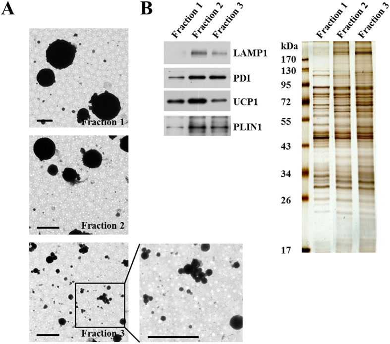
Figure 5. Separation of lipid droplet subpopulations from mouse brown adipose tissue by size.
LDs from mouse BAT were fractionated by size using differential centrifugation (Materials and Methods). Four LD fractions were collected. (A) EM to determine LD size. The four LD fractions were processed for positive staining and the sizes of the LDs were analyzed by EM. Bar = 2 μm. (B) Protein profile of the subpopulations. Proteins of LD subpopulations were extracted with chloroform:acetone (300 μl:700 μl) and analyzed by Western blotting with the indicated antibodies and silver staining (equal amount of protein per lane).

