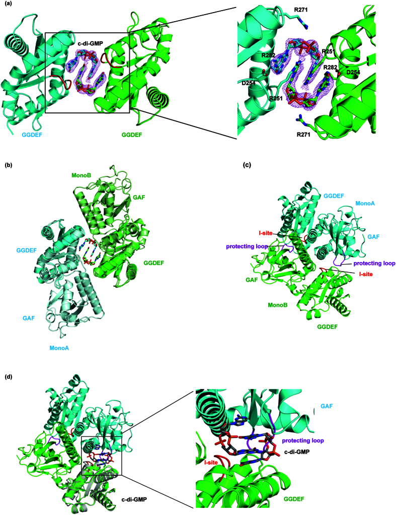
Figure 3. The structure of the GGDEF domain of Dcsbis in complex with c-di-GMP and the inhibition loop.
(a) The dimer of the Dcsbis-GGDEF/c-di-GMP complex. The I-site and A-site of Dcsbis are in red and blue, respectively. (b) The superposition of the full-length Dcsbis monomer onto the Dcsbis-GGDEF/c-di-GMP complex dimer. The full-length protein is in lighter colors. (c) The peptide loop (protecting loop, residues 120–125) extending from the GAF domain blocks the inhibition site of Dcsbis. The protecting loop is colored in purple, whereas the I-site is red and the active site is blue. (d) The superposition of GGDEF/c-di-GMP complex onto the full-length structure of Dcsbis to model the position of c-di-GMP into the full-length Dcsbis structure. GGDEF/c-di-GMP are highlighted in gray. A close view of the c-di-GMP modeled into the Dcsbis structure.

