Figure 6.
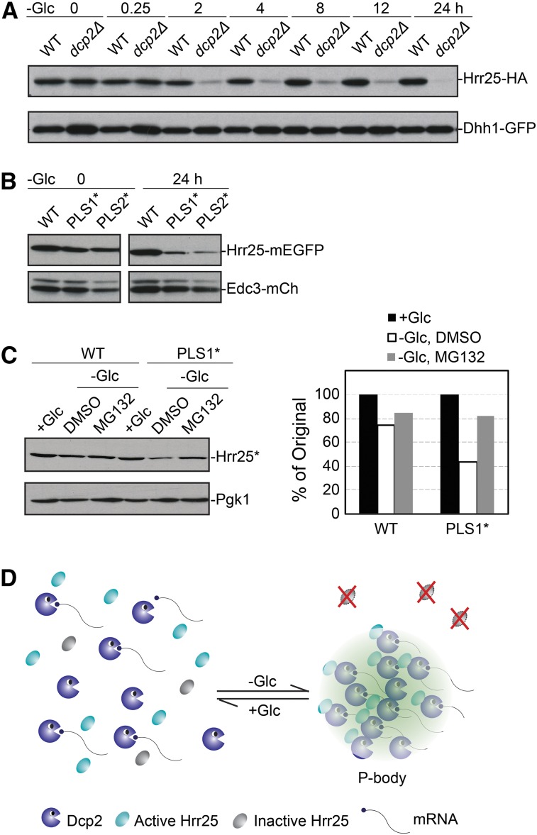
A failure to localize to P-bodies resulted in the rapid turnover of Hrr25. (A) Hrr25 protein levels significantly decreased in dcp2Δ cells upon glucose starvation. WT and dcp2Δ cells were starved for glucose for the indicated times and the levels of Hrr25 were then assessed by Western blotting. (B) Alterations of the PLS1 or PLS2 motifs resulted in the destabilization of Hrr25 upon glucose starvation. Strains expressing the indicated Hrr25 proteins were starved for glucose for 24 hr and the relative level of each protein was then assessed by Western blotting. (C) Inhibition of the proteasome stabilized the PLS1* variant upon glucose starvation. Strains expressing the indicated Hrr25 proteins were starved for glucose for 24 hr with or without the proteasome inhibitor, MG-132 (100 μM). The relative level of each protein was assessed by Western blotting (left) and quantified with the ImageJ software package (right). (D) A model showing how the localization to P-bodies sequesters Hrr25 away from the degradation machinery in the cytoplasm.

