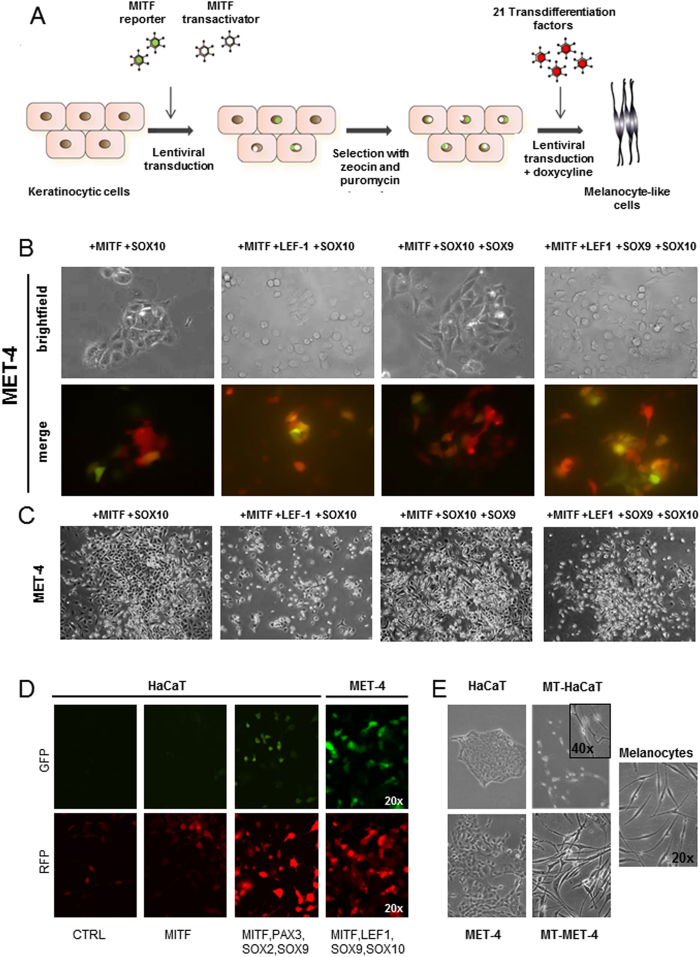
Figure 1. Transdifferentiation of HaCaT and MET-4 cells into melanocyte-like cells.
(A) Scheme of workflow. Cells were first selected for the M2 transactivator and MITF reporter and afterwards transduced with transcription factor cocktail and induced with doxycycline (B) Transgene expression was confirmed with different transcription factor combinations. Stronger RFP signal was observed depending on the amount of different factors used. Various transcription factor cocktails could induce MITF expression in MET-4 cells. The strongest expression was noted for the factor combination MITF, LEF-1, SOX9 and SOX10. (C) Different transcription factor combinations could induce morphological changes in MET-4 cells. The cells changed their morphology from cobblestone-like appearance into more spindle-shaped cells. Cell-cell adhesion was lost and colony formation was reduced. Single cells with loose contacts to the neighbouring cells were present. (D) Transgene expression (RFP) and MITF reporter activation (GFP) in HaCaT and MET-4 cells for different factor combinations (E) Morphology of HaCaT cells, MET-4 cells and melanocytes as well as morphological changes observed after induction of the transdifferentiation factor overexpression in melanocyte-transdifferentiated HaCaT (MT-HaCaT) and melanocyte-transdifferentiated MET-4 (MT-MET-4) cells.

