
Fig. 5.
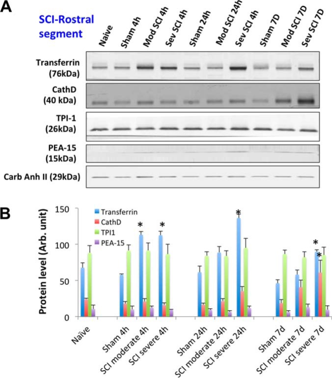
SCI-tissue immunoblotting validation (rostral segment). A, Time course of post-SCI biomarkers and new candidates validation illustrated by Western blot of rat spinal cord tissue (rostral) lysate of rostral section (sham, and two SCI severity) collected at three time points (4 h, 24 h, and 7 days), candidate markers probed include transferrin, cathepsin D, TPI-1 and PEA-15. Western blot of carbonic anhydrase II served as a loading control. B, Immunoblotting quantification of spinal cord tissue lysate samples (sham and two SCI severity levels at 3 time points) for transferrin, CathD, TPI-1, and PEA-15 biomarkers. Mean + S.E. are shown. (* p < 0.05, compared with corresponding sham, unpaired t test).