Fig. 1.
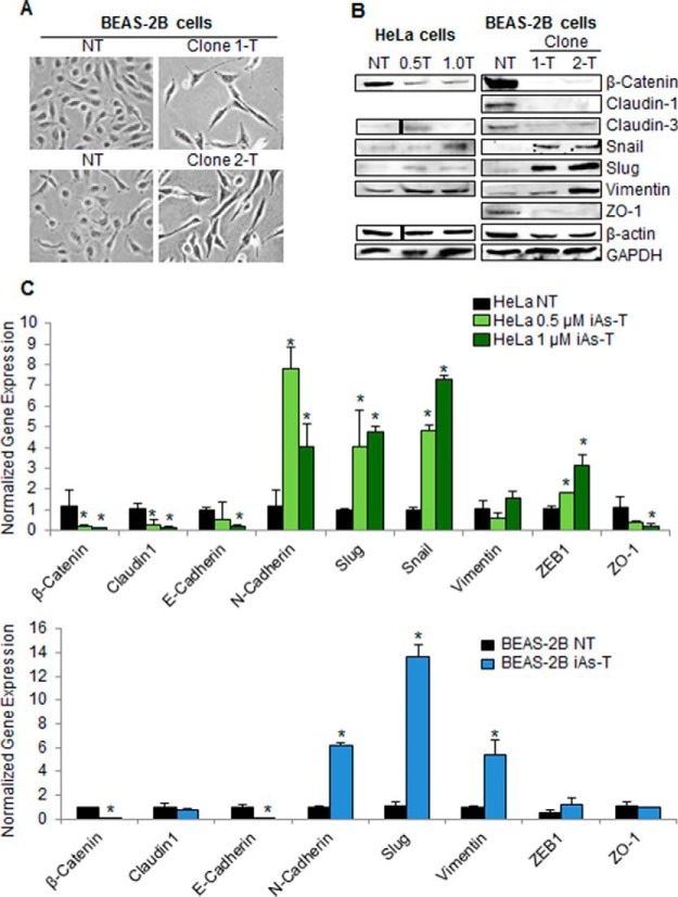
Chronic low-dose exposure to iAs causes cells to undergo EMT. A, Morphology changes seen in iAs-transformed BEAS-2B cells. BEAS-2B cells become elongated and spindle-shaped with 0.5 μm iAs treatment. B, Western blot analyses of EMT markers showing changes in the EMT markers. For instance, β-Catenin levels decrease and Slug levels increase in iAs transformed cells. C, qRT-PCR confirmation of iAs-induced EMT markers in HeLa (top) and BEAS-2B (bottom). qRT-PCR confirms the changes in expression of EMT markers in iAs-transformed cells. Error bars represent standard deviation (S.D.) from three biological replicates, each containing three technical replicates. Asterisks denote p values < 0.05 as determined by Student's t test.

