Fig. 1.

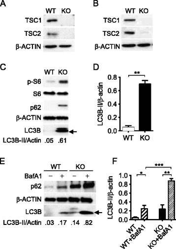
Loss of TSC1 led to sustained activation of the mTORC1 and to increased auto-phagosome formation. Representative Western blot of TSC1 and TSC2 in bone marrow macrophages from TSC1f/f-ERCre+ mice (a) and in the resident peritoneal macrophages of TSC1fl/fl LysM-Cre+ mice (b) did not detect the TSC1 and TSC2 proteins. Isolated cells from mice were grown in 15 % L929 conditional medium for 4 days. 4-Hydroxytamoxifen at 2.5 mM was added into L929 conditioned culture medium for 2–3 days before use and the protein lysates were prepared. c Immunoblot showed significantly increased basal p-S6, LC3B-II (arrow), and p62 proteins in macrophages isolated from TSC1 KO mice. d Densitometric quantification of LC3-II (n = 3) band intensities quantified by ImageJ was normalized to total Actin. e TSC1 WT and KO BMMϕ were treated with 100 nM bafilomycin A1 for 3 h to measure autophagic flux. f Densitometric quantification of LC3-II (n = 3) was normalized to total Actin. The experiments were repeated three times. The arrow indicates LC3B-II. BafA1: bafilomycin A1. *, p <0.05, **, p < 0.01, ***, p < 0.001
