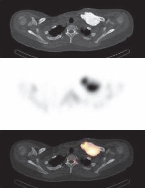
Figure 2.

The transaxial CT image (upper section) identifies a dense lesion with well-defined contours in the left clavicle. There is intensely increased uptake on the corresponding SPECT image (middle section) and SPECT/CT fusion image (lower section)
