Fig. 5.
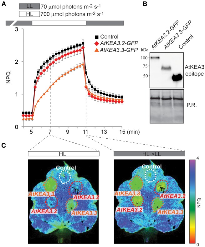
Overexpression of KEA3.2 and KEA3.3 in tobacco. (A) Chl fluorescence of tobacco leaves transformed with KEA3.2−GFP, KEA3.3−GFP and control (GFP fused to the KEA3 antibody-binding site coding sequence at its N-terminus) was monitored during alternating low light (70 μmol photons m−2 s−1, gray bar), high light (700 μmol photons m−2 s−1, white bar) and low light. Error bars represent the SEM (n = 4). (B) Proteins extracted from the transformed tobacco sections were immunodetected with the specific KEA3 antibody. Ponceau red (P.R.) staining of the membrane prior to immunodetection is shown as a loading control. (C) Images of a tobacco leaf transformed with KEA3.2−GFP, KEA3.3−GFP and control 120 s after transition from low to high light (HL) and 20 s after transition from high to low light (HL→LL 20 s) are shown. False colors represent the NPQ as indicated on the color scheme.

