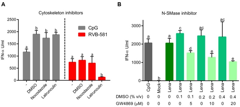
FIGURE 4.
Role of cytoskeleton and membrane ceramide in pDC activation by PRRSV-infected MØ. (A) MØ were infected with PRRSV RVB-581 for 90 min, washed and then treated with the cytoskeleton inhibitors nocodazole (10 μM; interferes with microtubule polymerization) or latrunculin (3 μM; inhibitor of actin polymerization) or DMSO as control for 2 h at 39°C. After three washes, MØ were co-cultured with pDC. Control wells were stimulated with CpG D32. DMSO controls showed an increase in IFN-α secretion by pDC stimulated with CpG D32. (B) MØ were infected with PRRSV Lena for 90 min, washed and then co-cultured with pDC. Then DMSO controls or different concentrations of GW4869 (inhibitor N-SMase) at 5, 10, and 20 μM were added to the co-cultures. Mock-treated MØ and CpG D32 stimulation were included as controls. N-SMases are important in the metabolism of sphingomyelin and required to produce cell membrane ceramide and phosphocholine, which are important during intercellular interactions. For (A,B), the IFN-α production was measured after 20 h of incubation in co-culture. The data represent mean values of three replicates with standard deviation, and represent three (A) and two (B) independent experiments. Different letters on top of the bars indicate significant difference based on the Mann–Whitney test (P < 0.05), whereas bars sharing same letters indicate no significant differences.

