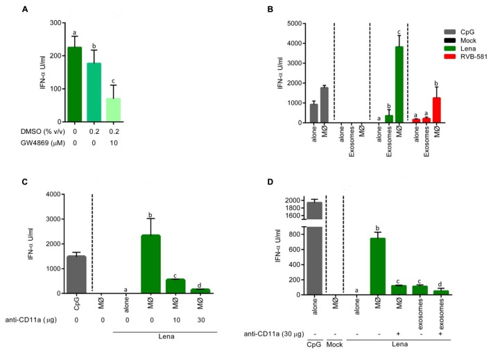
FIGURE 5.
pDC stimulation by PRRSV-infected MØ is dependent on integrin-mediated cell contact. (A) Stimulation of pDC by MØ-derived exosomes. MØ were infected with PRRSV Lena for 90 min followed by wash steps and addition of fresh medium. After 18 h of culture, exosome fractions were isolated and used to stimulate enriched pDC during 20 h at 39°C. The N-SMase inhibitor GW4869 was added at 10 μM, DMSO alone was added at 0.2% (v/v) or the cells were left untreated. (B) Stimulation of pDC by infected MØ is superior to exosome stimulation. pDC were stimulated with PRRSV (Lena or RVB-581) virions at MOI 0.1 TCID50/ml (“alone”), with PRRSV-infected (Lena or RVB-581) MØ, with exosome fractions isolated from supernatant of PRRSV-infected (Lena or RVB-581) MØ; CpG D32 was included as control of pDC activation. (C) pDC stimulation by infected MØ involves ITGAL (CD11a). pDC were stimulated with mock-treated MØ, with Lena virions at MOI 0.1 TCID50/ml (“alone”) and with Lena-infected MØ. Anti-CD11a mAb was added to the co-cultures at 10 or 30 μg/ml as indicated. CpG D32 was included as control for pDC activation. (D) Effect of anti-CD11a on pDC stimulation by infected MØ or exosomes. pDC were stimulated with mock-treated MØ, with Lena virions at MOI 0.1 TCID50/ml (“alone”), with Lena-infected MØ or with the exosome fraction isolated from Lena-infected MØ. Anti-CD11a mAb was added to the co-culture and to the exosome fraction stimulation at 30 μg/ml. CpG D32 was included as control for pDC activation. Experiments were done in triplicates, and represent three (A,B) or two (C,D) different experiments. Different letters on top of the bars indicate significant difference based on the Mann–Whitney test (P < 0.05), whereas bars sharing same letters indicate no significant differences.

