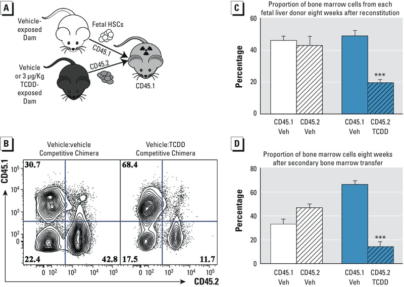
Figure 3.

Effects of transplacental tetrachlorodibenzo-p-dioxin (TCDD) exposure on the long-term self-renewal potential of fetal liver hematopoietic stem cells. (A) Schematic model of the experimental design for the primary reconstitution experiment. (B) Representative flow cytometry plot of bone marrow from the primary chimera. Numbers in each quadrant of the flow cytometry plots represent the percentage of bone marrow cells identified by the antibody specific for CD45.1 or CD45.2 congenic surface proteins. (C) Percent of bone marrow cells from each donor in the primary and secondary recipients. White bars represent control competitive chimeras, and blue bars represent the chimeras where vehicle (Veh) cells were competed with cells obtained from TCDD-exposed fetuses. Solid bars represent CD45.1+ cells, and CD45.2+ cells are denoted with diagonal slash-filled bars. Data are the mean ± SEM with 5 mice per group. The experiment was repeated twice. (D) Percent of bone marrow cells from each donor after the secondary bone marrow transfer. *** Indicates statistical significance by analysis of variance (ANOVA) followed by Tukey’s test; p < 0.01 compared with the congenic cells from the same chimera.
