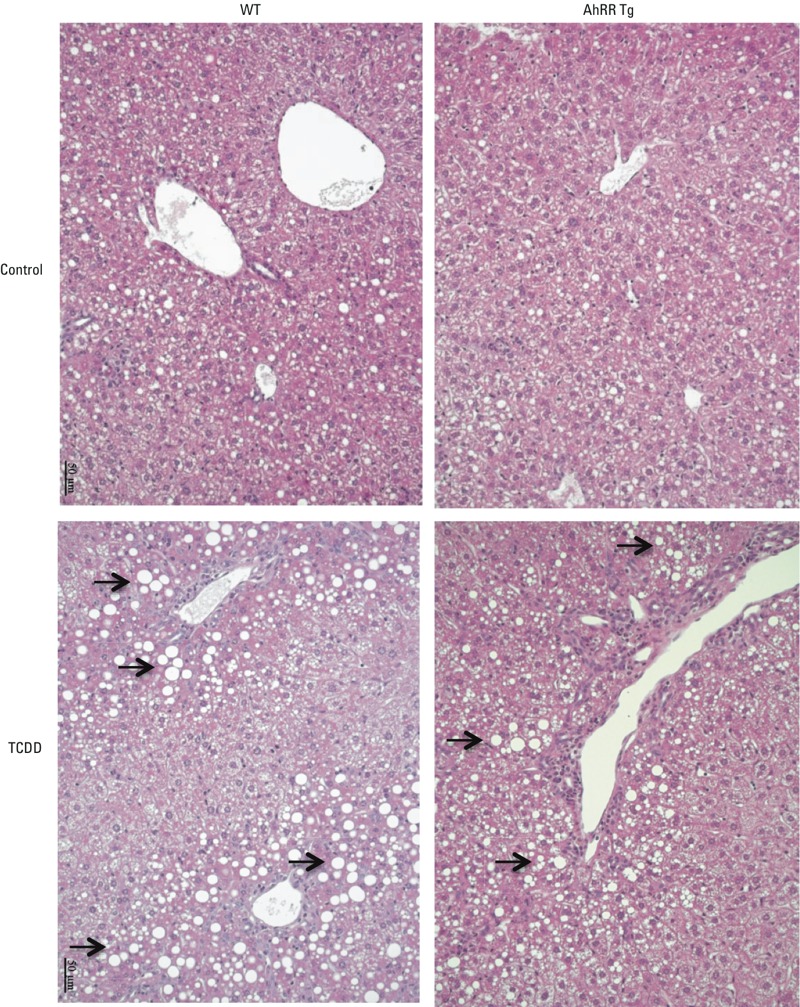
Figure 11.

Hematoxylin and eosin stained liver sections of male wt and AhRR Tg mice. Liver sections were prepared and stained following a single i.p. dose of 50 μg/kg TCDD for 6 days. The arrows indicate clear vacuoles caused by histological fixation dissolving the accumulated lipids. Images represent replicates from three mice in each group.
