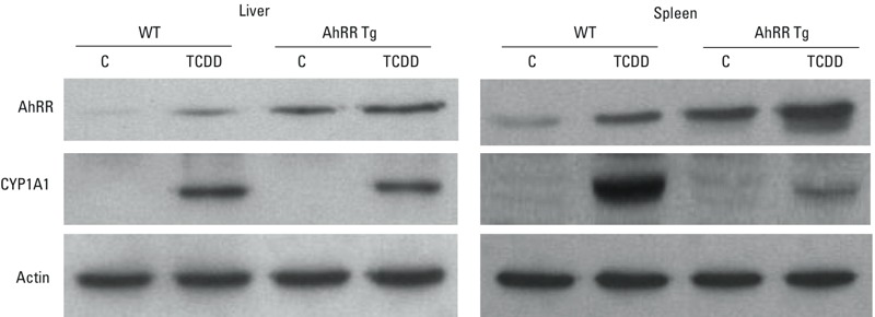
Figure 3.

Protein levels of AhRR and CYP1A1 in liver and spleen from male AhRR Tg and C57BL/6J wt mice. Proteins were isolated 24 hr after treatment with vehicle control (C) or 20 μg/kg TCDD and determined by Western blot analysis. Equivalent amounts of whole tissue protein (30 μg of protein) were loaded in each lane on 10% SDS-polyacrylamide gels and analyzed by immunoblotting using AhRR- and CYP1A1 specific antibodies. Bands of Western blot represent replicates from three mice in each group.
