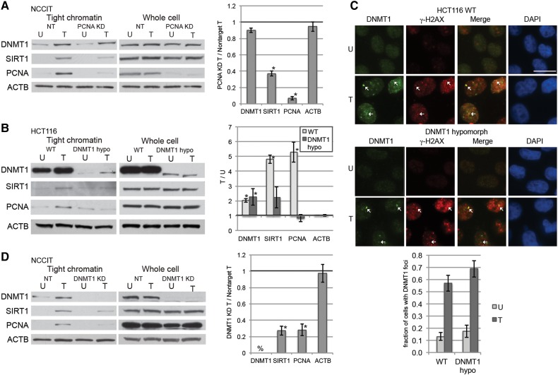
Figure 1.
Oxidative damage-induced recruitment of DNMT1 to chromatin is not dependent on PCNA. (A) NCCIT cells were transfected with nontarget siRNAs (NT) or PCNA siRNA. After 72 h, they were untreated (U) or treated with 1 mM H2O2 for 30 min (T). Data are displayed as mean ± SEM of the ratio of the indicated protein levels in PCNA-knockdown H2O2-treated cells over nontarget H2O2-treated cells (n = 2). *P < 0.05. (B) HCT116 parental (WT) or DNMT1 hypomorphic (DNMT1 hypo) cells were untreated (U) or treated with 4 mM H2O2 for 30 min (T). Data are displayed as mean ± SEM of the ratio of the indicated protein levels in H2O2-treated cells over untreated cells for the given cell line (n = 3). *P < 0.05. (C) HCT116 parental (WT) or DNMT1 hypomorphic (DNMT1 hypo) cells were untreated (U) or treated with 4 mM H2O2 for 30 min (T). Immunofluorescence analysis was performed after using a preextraction buffer. Arrows indicate foci where DNMT1 and γ-H2AX colocalize. Data presented are mean ± SEM of the percentage of cells with DNMT1 foci in their nuclei from three biological replicates with at least 20 cells counted for each replicate. Scale bar, 5 μm. (D) NCCIT cells were infected with nontarget shRNAs (NT) or DNMT1 shRNA. After 96 h, they were untreated (U) or treated with 1 mM H2O2 for 30 min (T). Data are displayed as mean ± SEM of the ratio of the indicated protein levels in DNMT1-knockdown H2O2-treated cells over nontarget H2O2-treated cells (n = 3). *P < 0.05. %, DNMT1 is not detectable by western blot.

