
Figure 1.
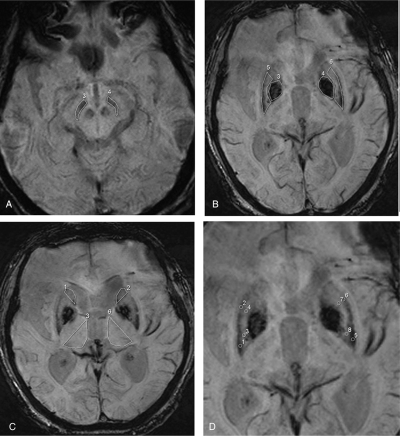
Axial susceptibility-weighted magnetic resonance imaging illustrating the region of interest placements in the (A) substantia nigra in the midbrain, (B) putamen (numbered 5,6) and globus pallidus (numbered 3,4), (C) head of caudate nucleus (numbered 1,2) and thalamus (numbered 3,6), and the 4 subregions of the putamen (D).