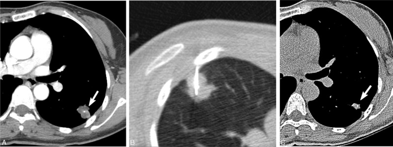
Figure 2.

A 73-year-old asymptomatic woman with an SPN. A, CT showed a lobulated nodule (arrow) with poor contrast enhancement (HU, 41) in the left lobe. Note the internal dot-like calcification. B, CT-guided PCNB using a 20-gauge Westcott needle showed chronic granulomatous inflammation. Culture of PCNB aspirates showed the presence of Mycobacterium avium. PCNB aspirates were negative for tuberculosis-PCR and positive for NTM-PCR. C. Follow-up CT obtained 2 years later without anti-MAC antibiotic treatment showed a decrease in the size of the NTM nodule (arrow). CT = computed tomography, MAC = Mycobacterium avium, NTM = nontuberculous mycobacterial, PCNB = percutaneous needle aspiration biopsy, PCR = polymerase chain reaction.
