
Figure 3.
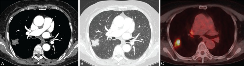
A 73-year-old asymptomatic woman with an SPN. A, Contrast-enhanced CT shows a lobulated nodule with poor contrast enhancement with a mean attenuation value of 33 HU. B, Lung window CT image showed a nodule with a lobulated contour in the right upper lobe. No abnormal findings such as bronchiectasis or clustered small nodules in the remaining lungs were noted. C, Fused axial image from 18F-FDG PET/CT showed strong FDG uptake (SUVmax, 7.8). This patient underwent surgical resection (right upper lobectomy) without PCNB. Surgical specimen showed chronic granulomatous inflammation and was positive for NTM-PCR. D, Fused axial image. CT = computed tomography, FDG = fluorodeoxyglucose, MAC = Mycobacterium avium, NTM = nontuberculous mycobacterial, PCNB = percutaneous needle aspiration biopsy, PCR = polymerase chain reaction, SUVmax = maximum standardized uptake value.