
Figure 4.
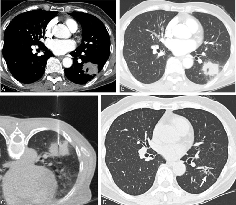
A 61-year-old asymptomatic woman with a mass-like consolidation. A, Contrast-enhanced CT showed a mass-like consolidation with poor contrast enhancement with a mean attenuation value of 39 HU. B, Lung window CT image showed an irregular border and ill-defined ground-glass opacity adjacent to the mass-like consolidation. C, CT-guided PCNB using a 22-gauge Westcott needle showed chronic granulomatous inflammation. Culture of PCNB aspirates revealed the presence of Mycobacterium intracellulare organisms. D, Follow-up CT obtained 2 years after anti-MAC antibiotic treatment showed complete resolution with minimal fibrotic change (arrow). CT = computed tomography, MAC = Mycobacterium avium, PCNB = percutaneous needle aspiration biopsy.