Figure 3.

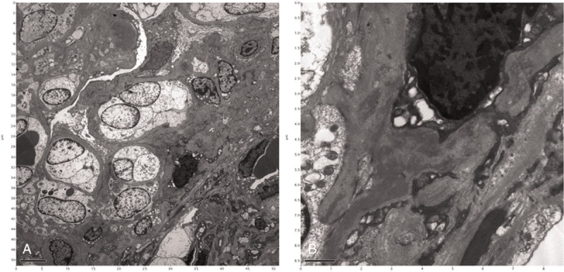
Electron microscopy demonstrating (A) expansion of the mesangial matrix (original magnification ×4000) and (B) electron-dense deposits in the mesangium (original magnification ×20,000).

Electron microscopy demonstrating (A) expansion of the mesangial matrix (original magnification ×4000) and (B) electron-dense deposits in the mesangium (original magnification ×20,000).