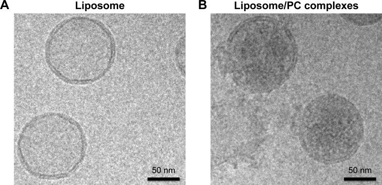
Figure 1.
Liposome and liposome/PC complex characterization by cryo-electron microscopy.
Notes: High-magnification cryo-EM images of liposomes before (A) and after incubation in plasma (B). Cryo-EM analysis reveals a spherical and unilamellar shape for both samples. Liposomes retained their shape and structure after incubation with plasma. Note the significant difference in electron density on the particle surface after plasma incubation, which indicates the presence of the PC (B).
Abbreviations: PC, protein corona; cryo-EM, cryo-electron microscopy.

