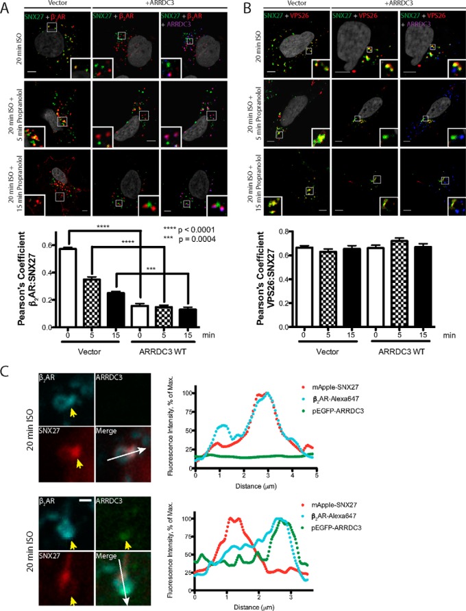
FIGURE 6.
Overexpression of ARRDC3 decreases the co-localization of the β2AR and SNX27 on endosomes. A, representative images and corresponding quantifications of β2AR localization with endogenous SNX27 in cells with or without ARRDC3 overexpression were analyzed by immunofluorescence staining upon 20 min of treatment with 10 μm ISO followed by 10 μm propranolol treatment for the indicated times. Three independent experiments were performed. Pearson's coefficients are the mean ± S.E. and compared using an unpaired t test (n = 30). Scale bars, 5 μm. B, representative images and corresponding quantifications of the localization of endogenous SNX27 and VPS26 in cells with or without ARRDC3 overexpression were analyzed by immunofluorescence staining upon 20 min of treatment with 10 μm ISO followed by 10 μm propranolol treatment for the indicated times. Three independent experiments were performed. Pearson's coefficients are the mean ± S.E. and compared using an unpaired t test (n = 30). Scale bars, 5 μm. C, representative immunofluorescence confocal live cell images and corresponding line scan analysis are shown to demonstrate the localization of the β2AR, pEGFP-ARRDC3, and mApple-SNX27 on the same endosomes. FLAG-β2AR-stable HEK293 cells were transiently transfected with mApple-SNX27 and vector/pEGFP-ARRDC3. Three independent experiments were performed, and over 50 endosomes were examined. Scale bars, 1 μm.

