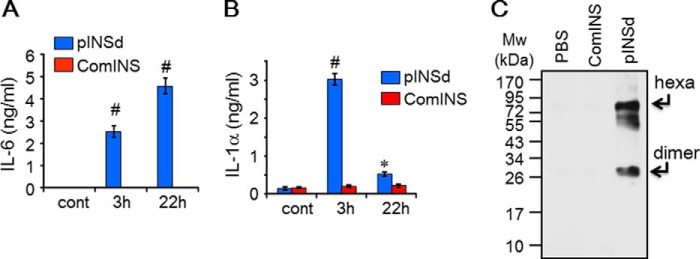
FIGURE 2.
Detection of a common motif in both IL-1α and pINS. IL-6 levels in the supernatant of A549 cells treated with pINSd were increased by prolonged incubation (A), but IL-1α levels were decreased with prolonged incubation (B) as measured by the IL-1α ELISA kit described under “Experimental Procedures.” Incubation time is indicated on the x axis. C, Western blot with affinity-purified rabbit polyclonal antibody raised against IL-1α detected 26 (dimer)- and 75 (hexamer (hexa))-kDa size bands where pINSd was added but not where PBS or comINS was added. Data in A and B are comparisons between pINSd treatment and untreated control (cont). Data are mean ± S.E. (error bars). *, p < 0.05; #, p < 0.001 from duplicates.

