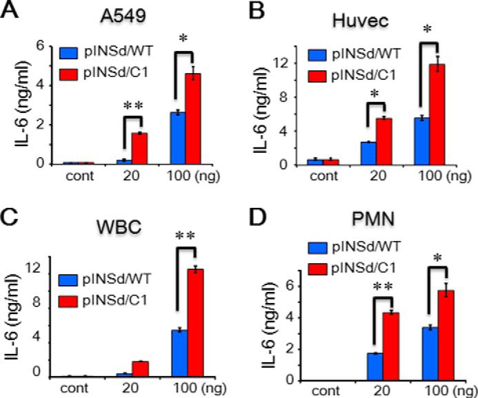
FIGURE 6.

Bioassay of the motif-deleted pINSd mutant. A–D, assay of pINSd/WT and /C1. A549 cells (A), Huvecs (B), WBCs (C), and PMN leukocytes (D) were treated with pINSd/WT and /C1. The INS/IL-1α motif-deleted pINSd/C1 mutant was more active than pINSd/WT across different cell type. Data in A–D are comparisons between pINSd/WT and /C1 mutant. Data are mean ± S.E. (error bars). *, p < 0.05; **, p < 0.01 from duplicates. cont, control.
