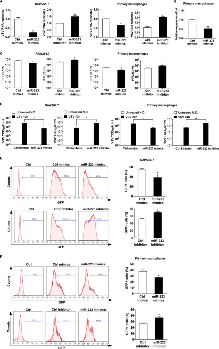
FIGURE 3.
miR-223 feedback attenuates the viral replication. A, RAW264.7 cells transfected with miR-223 mimics or inhibitors were infected by VSV at m.o.i. 0.1 for 1 h, then washed, and fresh medium was then added; after 12 h, VSV intracellular VSV RNA replicates were quantified using qRT-PCR and normalized to that of β-actin in each sample. Mouse peritoneal macrophages transfected with miR-223 mimics or inhibitors were infected by VSV at m.o.i. 10 for 1 h, then washed, and fresh medium was then added; after 24 h, intracellular VSV RNA replicates were quantified using qPCR and normalized to that of β-actin in each sample. B, mouse peritoneal macrophages transfected with miR-223 mimics were infected by H1N1 at m.o.i. 10 for 1 h, then washed, and fresh medium was then added; after 24 h, intracellular HA mRNA was quantified using quantitative RT-PCR. C and D, RAW264.7 and peritoneal macrophages were treated as in A, and VSV Plaque assay and TCID50 assay in cultural supernatants were done on Vero cells (N.D., not detected). E and F, RAW264.7 cells transfected with miR-223 mimics or inhibitors were infected by VSV-GFP at m.o.i. 0.1 for 1 h, then washed, and fresh medium was then added; after 12 h, VSV-GFP infection was quantified using flow cytometry. Mouse peritoneal macrophages transfected with miR-223 mimics or inhibitors were infected by VSV-GFP at m.o.i. 10 for 1 h, then washed, and fresh medium was then added; after 24 h, VSV-GFP infection was quantified using flow cytometry. Data are the mean ± S.D. (n = 3) of one representative experiment. Similar results were obtained in three independent experiments. **, p < 0.01; *, p < 0.05; Ctrl, control.

