FIGURE 4.
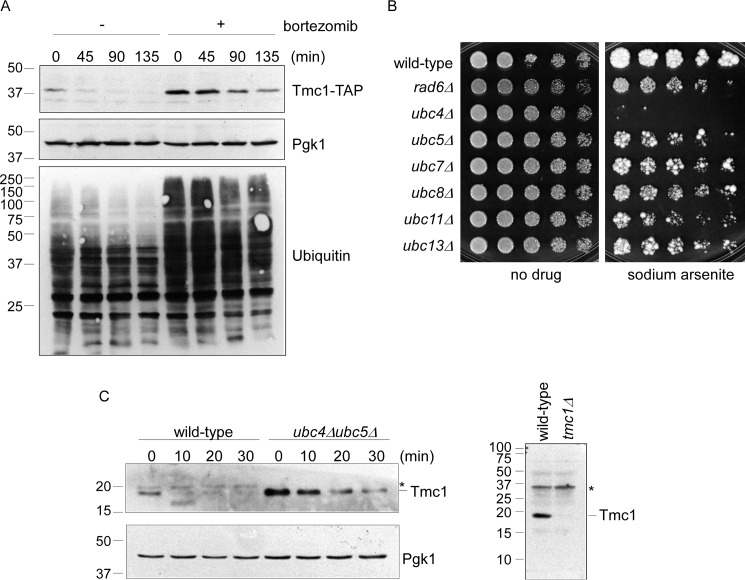
Tmc1 is a short-lived substrate of the ubiquitin-proteasome pathway. A, cycloheximide chase analysis of Tmc1 turnover in the presence or absence of the proteasome inhibitor bortezomib (30 μm). Whole cell extracts were prepared and analyzed by SDS-PAGE followed by immunoblot. Top panel, anti-TAP antibody. Middle panel, anti-Pgk1 antibody (loading control). Bottom panel, anti-ubiquitin antibody, which confirms robust inhibition of the proteasome after bortezomib treatment. B, phenotypic analysis of seven E2 ubiquitinating enzyme mutants. The indicated strains were spotted in 3-fold serial dilutions and grown in the presence or absence of sodium arsenite (1.5 mm) for 1.5–5 days at 30 °C. C, cycloheximide chase analysis of Tmc1 in wild-type cells and a ubc4Δubc5Δ double mutant. Left panels, whole cell extracts were prepared and analyzed by SDS-PAGE followed by immunoblot with anti-Tmc1 and anti-Pgk1 (loading control) antibodies. Note that endogenous (left panel) and TAP-tagged (A) Tmc1 show similarly short half-lives. Right panel, validation of the Tmc1 antibody. Whole cell extracts from wild-type and tmc1Δ strains were prepared and analyzed by SDS-PAGE followed by immunoblot with anti-Tmc1 antibody. Asterisk, nonspecific immunoreactive band.

