FIGURE 1.
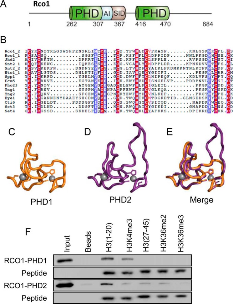
The PHD fingers of Rco1 bind to the extreme N terminus of H3. A, a schematic representation of the Rco1 protein. PHD fingers are highlighted in green with the autoinhibitory domain (AI) in blue and the Sin3 interaction domain (SID) in gray. B, an alignment of yeast PHD fingers, highlighting the conserved cysteine and histidine residues. C, a molecular model of PHD1. Zinc atoms are in gray. D, a molecular model of PHD2. Zinc atoms are in gray. E, a merge of the models of PHD1 and PHD2 show a high degree of similarity. F, in-solution peptide pulldown assays with PHD1 and PHD2 were carried out with the indicated histone H3 peptides. Both domains bind the unmodified H3 N terminus and show sensitivity to H3K4me3.

