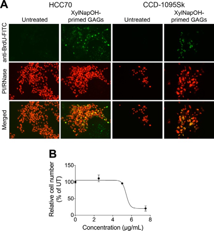
FIGURE 4.

XylNapOH-primed GAGs from HCC70 cells induce apoptosis of HCC70 cells and CCD-1095Sk cells. A, HCC70 cells (left panels) and CCD-1095Sk cells (right panels) untreated or treated with 5 μg/ml XylNapOH-primed GAGs from HCC70 cells for 24 h. DNA strand breaks indicating apoptosis were detected utilizing BrdU and visualized using an anti-BrdU-FITC antibody. Cell nuclei were stained using propidium iodide (PI)/RNase. The merged images displays colocalization of anti-BrdU-FITC-positive cells and cell nuclei. The images are representative for each experiment, performed at least in duplicate. B, HCC70 cells treated with XylNapOH-primed GAGs at the indicated concentrations for 24 h. The experiment was performed in duplicate in which n = 3. The data points are the means ± S.D.
