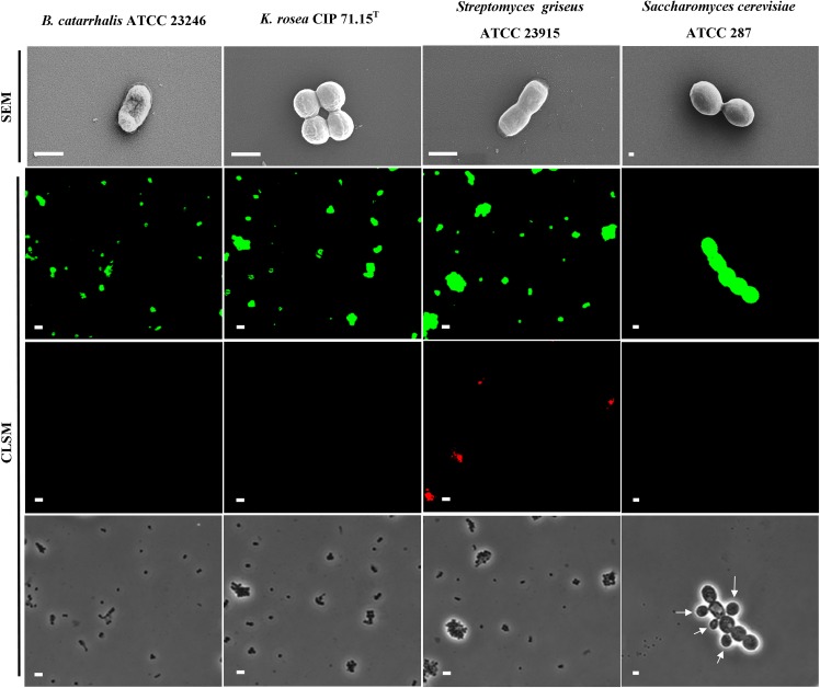
Fig 3. The effects of 18 GHz EMF radiation on the cells under investigation.
Typical SEM micrographs of B. catarrhalis, K. rosea, Streptomyces griseus bacteria and Saccharomyces cerevisiae yeast cells after exposure to 18 GHz EMF radiation. No significant change in cell morphology was observed. Scale bars 1 μm (top row). CLSM images show an uptake of the 23.5 nm nanospheres (second row) and 46.3 nm nanospheres (third row), after cell walls were permeabilised as a result of EMF exposures. The phase contrast images (in the bottom row) show the cells in the same field. Arrow indicates young yeast cells, which were unable to uptake any of the nanosheres. Scale bars 5 μm (second, third and bottom rows).

