Figure 1.
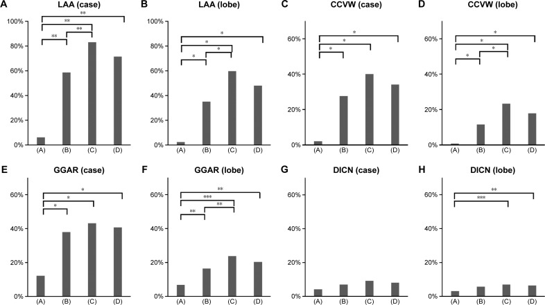
Incidence of thin-section computed tomography findings in nonsmokers and smokers (172 cases and 1,032 lobes).
Notes: Incidence of LAA in cases (A), LAA in lobes (B), CCVW in cases (C), CCVW in lobes (D), GGAR in cases (E), GGAR in lobes (F), DICN in cases (G), DICN in lobes (H). The incidence of LAA, CCVW, and GGAR was significantly higher in smokers than in nonsmokers in both case-based and lobe-based analyses. By lobe-based analysis, heavy smokers manifested a significantly higher incidence of CCVW and GGAR than moderate smokers. By lobe-based but not case-based analysis, the incidence of DICN was significantly higher in smokers than nonsmokers. (A) Nonsmokers, (B) mild/moderate smokers, (C) heavy smokers, and (D) all smokers. *P<0.001, **P<0.005, ***P<0.05.
Abbreviations: LAA, low attenuation area; CCVW, clustered cysts with visible walls; GGAR, ground-glass attenuation with/without reticulation; DICN, diffuse ill-defined centrilobular nodules.

