Figure 3.
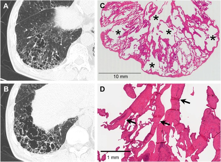
Severe SRIF with emphysema pattern on thin-section CT images and histological findings.
Notes: (A and B) Thin-section CT images showing clustered cysts of markedly irregular size and shape accompanied by ground-glass attenuation with reticular structures in the surrounding area. (C) Low-power photograph of a histological section (hematoxylin–eosin stain) reveals irregularly shaped emphysematous spaces (asterisk) with collagenous fibrotic walls. Many of the walls are truncated. Patchy fibrosis is observed in the intervening lung parenchyma and corresponds to ground-glass attenuation with reticular structures on thin-section CT. Irregular cysts with thickened walls tend to be present a little apart from the pleura with less-involved subpleural lung parenchyma. (D) On this high-power photograph of a histological section, fibrosis consists of hyalinized paucicellular fibrosis (arrow) corresponding to SRIF.
Abbreviations: SRIF, smoking-related interstitial fibrosis; CT, computed tomography.

