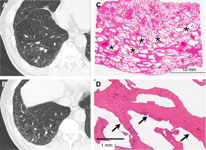
Figure 4.
Mild SRIF with emphysema pattern on thin-section CT images and histological findings.
Notes: (A and B) Thin-section CT images showing clustered cysts with visible walls in the peripheral zone with less involvement of the subpleural parenchyma. The size and shape of the cysts vary. Note ground-glass attenuation with reticular or branching structures in the surrounding area. (C) This low-power photograph of the histological section (hematoxylin–eosin stain) reveals irregularly shaped emphysematous spaces (asterisk) with thickened fibrotic walls corresponding to clustered cysts with visible walls on the thin-section CT image. There is patchy fibrosis at peribronchiolar and subpleural sites corresponding to ground-glass attenuation with reticular or branching structures in the surrounding area. Irregular cysts with thickened walls are present apart from the pleura with less-involved subpleural lung parenchyma. (D) On this high-power photograph of a histological section, fibrosis of this pattern consists of hyalinized paucicellular fibrosis (arrow) or SRIF.
Abbreviations: SRIF, smoking-related interstitial fibrosis; CT, computed tomography.

