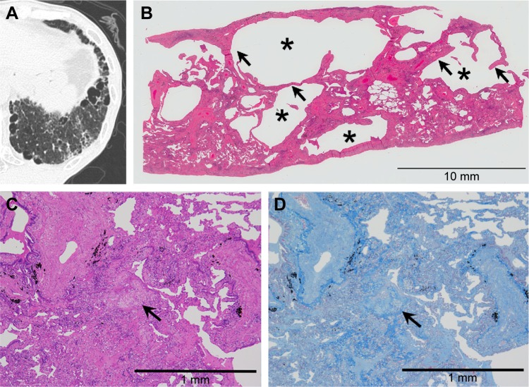
Figure 6.
Combined SRIF and UIP patterns on thin-section CT images and histological findings.
Notes: (A) Thin-section CT images showing foci of clustered cysts of irregular size and shape and surrounding ground-glass attenuation with reticular or branching structures. (B) This low-power photograph of the histological section (hematoxylin–eosin stain) reveals areas with irregularly shaped emphysematous spaces (asterisk) and hyalinized fibrotic walls (arrow). (C) High-power photograph of a histological section. Note dense fibrosis and fibroblastic foci (arrow) in the same histological specimen. (D) Masson’s trichrome stain reveals architecture destruction with irregularly distributed dense fibrosis. The fibroblastic focus (arrow) is seen inside the fibrosis.
Abbreviations: SRIF, smoking-related interstitial fibrosis; UIP, usual interstitial pneumonia; CT, computed tomography.

